The word “microplasty” can describe different knee procedures, from marrow-stimulation microfracture for a focal cartilage defect to a surgeon-specific resurfacing technique. You can judge whether the term fits your symptoms by matching the pain pattern, imaging, knee stability, alignment, and treatment history before discussing an operation.
Key takeaways
- Ask your surgeon whether microplasty means microfracture or focal implant resurfacing.
- Confirm localized damage with an examination, weight-bearing X-rays, and MRI when needed.
- Microplasty suits selected small lesions, not widespread arthritis or major knee instability.
- Compare recovery, durability, and rehabilitation before choosing microplasty over other treatments.
What does “microplasty” mean in knee surgery?
“Microplasty” is not a universally standardised knee-surgery name. The microplasty meaning depends on the surgeon: it often describes microfracture, but some use it for resurfacing a single damaged area with an implant.
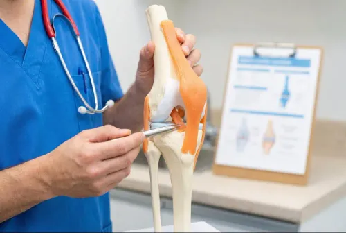
The treated problem is usually a focal cartilage defect: a full-thickness loss of articular cartilage on the medial or lateral femoral condyle, patella, or trochlea. It is different from arthritis wearing down an entire compartment.
For microfracture knee surgery, the surgeon:
- Removes loose or unstable cartilage and trims the edges into a stable rim.
- Makes controlled holes through the subchondral bone beneath the defect.
- Allows marrow elements to form a repair clot over the exposed surface.
No implant is inserted. The clot supports fibrocartilage repair, which does not reproduce normal hyaline cartilage.
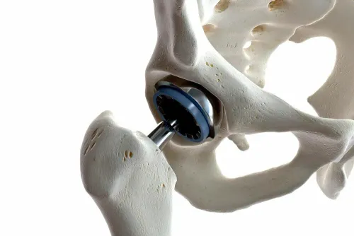
If “microplasty” means covering the damaged area with metal and polyethylene, ask whether the operation is actually a focal resurfacing implant, a surgeon-specific technique, or a unicompartmental knee replacement (UKR). A standard UKR replaces the femoral and tibial surfaces of one entire medial or lateral tibiofemoral compartment.
Microfracture treats a cartilage defect without replacing that compartment.
The label alone cannot tell you the implant, durability, risks, or rehabilitation plan. Request the exact procedure name, treated surface, implant details, and operative objective before deciding.
How is localized knee damage confirmed?
Doctors confirm localized knee damage by matching your symptoms and examination findings to imaging that shows one affected area—not merely by finding a cartilage abnormality.
- Pain location, swelling, catching, locking, instability, and walking tolerance
- Range of motion, tenderness, knee alignment, ligament stability, and patellar tracking
- Meniscal signs, plus possible hip or spine causes of referred pain
- A weight-bearing knee X-ray, taken while standing, to show joint-space narrowing under load
- Anteroposterior and lateral views, plus a skyline or Merchant view for the patellofemoral joint
- A flexion posteroanterior view, such as a Rosenberg view, when early narrowing is difficult to see
- MRI when cartilage thickness, an MRI cartilage defect, meniscus tissue, bone-marrow lesions, or ligament condition remains unclear after examination and X-rays
Doctors map the finding to one of three compartments: the medial tibiofemoral compartment, lateral tibiofemoral compartment, or patellofemoral compartment. A patella or trochlea defect requires particular attention to tracking and instability; treating cartilage alone will not correct abnormal kneecap loading.
Imaging must match the pain pattern. An incidental MRI defect does not prove that defect causes symptoms, and an apparently filled defect after microfracture does not guarantee normal mechanical function. Persistent pain can instead arise from meniscal damage, malalignment, bone-marrow lesions, ligament problems, or disease in another compartment.
If more than one compartment is diseased, treating one surface will not correct pain generated elsewhere.
Which patients are reasonable candidates, and who is a poor fit?
A reasonable microfracture candidate has a symptomatic, focal, contained, full-thickness cartilage defect with healthy surrounding cartilage. Many surgeons discuss a cartilage lesion size of about 2 cm² or less, but no universal cutoff exists.
Use this practical checklist:
- Preserved cartilage in the other knee compartments
- Acceptable knee alignment without uncorrected varus or valgus loading
- Useful range of motion without substantial stiffness
- Ligament stability, including intact or adequately functioning ACL, PCL, and collateral ligaments
- Symptoms that match the lesion rather than another compartment, meniscus, or bone problem
- A review of body weight, age as a risk modifier, work, sport, and impact demands
- Inadequate improvement after activity modification, quadriceps-and-hip physiotherapy, weight reduction when relevant, analgesics, safe anti-inflammatory medication, bracing, or an injection programme
| Feature | More suitable | Poor fit |
|---|---|---|
| Lesion | Small, contained, focal defect | Large, uncontained, or degenerative defect |
| Joint condition | Other compartments preserved | Diffuse osteoarthritis or substantial joint-space narrowing |
| Mechanics | Correctable alignment and stable ligaments | Severe deformity, uncorrected malalignment, or instability |
| Tissues | Adequate meniscus and bone | Important meniscal deficiency or major bone loss |
| Health | No infection or inflammatory arthritis | Infection or inflammatory arthritis |
Larger defects, older age, higher body weight, long-standing symptoms, degenerative disease, and high-impact goals make fibrocartilage repair less predictable. Bone-marrow stimulation does not reliably recreate normal hyaline cartilage, so it is not a general treatment for wear-and-tear arthritis.
How does microplasty compare with other treatments?
Before choosing surgery, compare microfracture with a completed non-operative knee treatment programme and with procedures that address the cause of abnormal loading. Try temporary activity modification, structured quadriceps-and-hip strengthening, weight reduction when appropriate, pain relief, and anti-inflammatory medication when safe. An unloading brace suits selected alignment patterns, while a corticosteroid injection offers short-term symptom control.
PRP injection and other biologic injections have different evidence and do not reliably rebuild worn cartilage. Reassess surgery when pain, swelling, or lost function persists despite the programme—not simply because an injection failed.
| Option | Main advantage | When it fits |
|---|---|---|
| Microfracture | Lower procedural burden; no graft harvest or implant | Small, contained focal defects with stable surrounding structures |
| Osteochondral autograft or allograft | Replaces the defect with bone and cartilage | Larger or high-demand focal defects |
| Cell-based cartilage restoration | Uses cartilage cells or tissue-engineering methods | Selected larger defects after specialist assessment |
| Total knee replacement | Treats multiple damaged compartments | Arthritis spans compartments or pain is not confined to one area |
A meniscus tear and cartilage defect may be treated during compartment-preserving surgery, but a large unreparable tear or meniscal deficiency leaves abnormal loading. ACL or PCL injury may require reconstruction first, and patellar maltracking may require realignment or stabilisation rather than microfracture alone.
Use a total knee replacement comparison when deformity is substantial, ligaments are unreliable, or arthritis is widespread. Compare rehabilitation time, implant longevity, activity limits, and possible revision—not just the operation itself.
What should you expect from the decision and recovery process?
Progressive activity-related pain, swelling, catching, or reduced function deserves evaluation when examination and imaging suggest one focal lesion. Seek urgent assessment if you cannot bear weight, have fever with a hot swollen knee, or develop sudden calf swelling and breathlessness.
1. Confirm the target: identify the exact compartment, defect size and depth, alignment, radiographic findings, menisci, and ACL, PCL, and collateral ligaments. An incidental MRI defect does not prove it causes your pain.
2. Prepare for surgery: arrange blood tests, anaesthetic checks, medication and anticoagulant instructions, transport, home support, and time away from work. Ask whether a combined meniscus, ligament, or osteotomy procedure changes the plan.
3. Plan knee surgery recovery: expect early controlled movement, wound care, physiotherapy, stair practice, and possibly crutches with restricted weight bearing for several weeks. Microfracture rehabilitation varies with lesion site, size, depth, and combined procedures; driving and impact exercise may remain restricted for months.
4. Weigh the risks of knee surgery: discuss infection, blood clots, stiffness, persistent pain, anaesthetic complications, instability, implant loosening or wear, arthritis in another compartment, and revision to total knee replacement.
Useful orthopaedic consultation questions include:
- Which named operation is proposed, and does it use an implant?
- What milestones determine weight bearing, driving, work, and sport?
- What follow-up and revision pathway applies?
Dr. Prajyot Jagtap can apply this framework when matching robotic, joint-preserving, regenerative, or reconstructive treatment to examination and imaging findings.
Related service
Joint Preservation Microplasty Microplasty is an advanced joint preservation procedure designed for patients with early-stage joint damage or partial cartilage wear. |
Frequently asked questions
What does “microplasty” mean in knee surgery?
Microplasty is not a universally standardised term. A surgeon may use it for microfracture, which creates small holes in bone beneath damaged cartilage, or for resurfacing one damaged area with an implant. Ask which exact procedure, device, and goal they mean.
How is localized knee damage confirmed?
Confirmation usually combines your symptoms, a physical examination, weight-bearing knee X-rays, and an MRI when cartilage, meniscus, ligaments, or a small bone lesion needs closer assessment. The surgeon must also check alignment and the condition of the rest of the knee.
Who is a reasonable candidate for microplasty?
A reasonable candidate has a clearly defined, limited damaged area, suitable knee alignment, stable ligaments, and little or no widespread arthritis. The decision also depends on age, activity goals, previous surgery, body weight, and the specific procedure proposed.
Who is a poor fit for microplasty?
Microplasty is a poor fit when arthritis affects multiple compartments, the knee has substantial deformity or instability, the lesion is too large or unsuitable, or symptoms do not match the scan. A surgeon must address these factors before recommending focal treatment.
How does microplasty compare with other treatments?
Options can include physiotherapy, activity modification, medicines or injections, microfracture, osteochondral grafting, autologous cartilage procedures, partial knee replacement, or total knee replacement. The comparison should cover lesion size, joint health, expected durability, risks, and rehabilitation time.