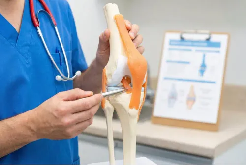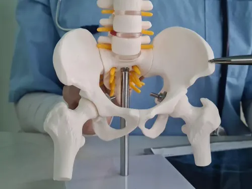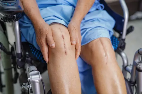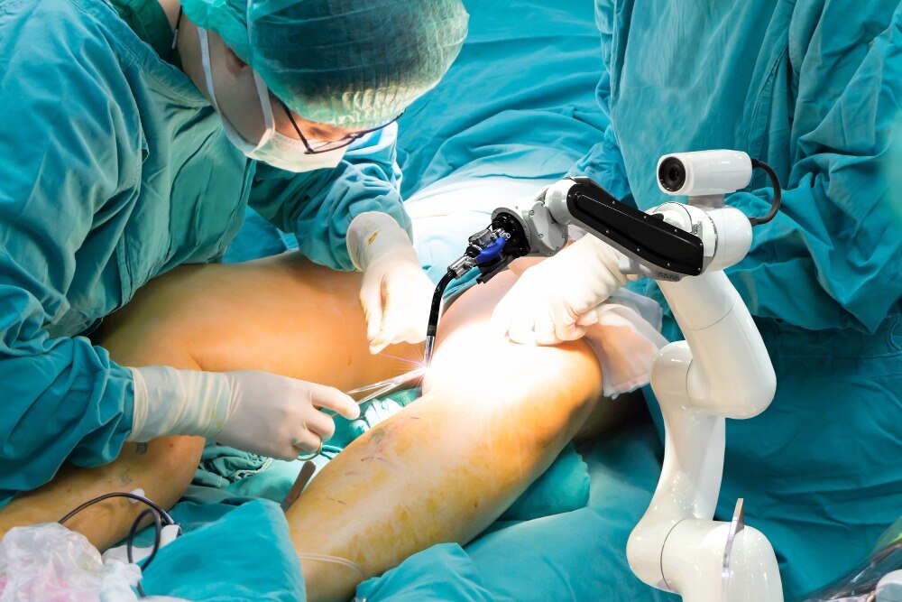
A successful knee replacement depends on more than placing an implant in a straight-looking leg: the plan must account for bone shape, deformity, ligament tension, stability, motion and implant position. By the end, you will know how alignment is planned and checked, what the robotic system controls, what remains the surgeon’s responsibility, and which details to ask about before consenting.
Key takeaways
- Alignment positions the femoral and tibial components for balanced knee movement.
- Preoperative CT or imaging creates the patient-specific alignment plan.
- The robot guides bone preparation but the surgeon controls each surgical step.
- Compare planning methods, surgeon experience, implant design, and verification protocols.
What joint alignment means in a knee replacement
Joint alignment in knee replacement describes how the femoral and tibial components sit in relation to each other and to the rest of the leg.
A straight line from the centre of the hip to the centre of the ankle represents the limb’s mechanical axis, but that line does not fully describe whether the knee will feel stable or move naturally.
Surgeons assess alignment in several directions:
- Coronal alignment: the inward or outward angle seen from the front, including varus “bow-legged” and valgus “knock-kneed” positioning.
- Sagittal alignment: the position viewed from the side, including the slope of the tibial component and the knee’s flexion or extension.
- Rotational alignment: how the femoral and tibial components turn around the limb, affecting kneecap tracking and movement.
- Mechanical alignment: positioning intended to bring the hip-knee-ankle axis close to a planned straight line.
- Anatomical alignment: positioning that follows the patient’s bone shape and native joint-line orientation rather than forcing every knee to the same straight-axis target.
Traditional mechanically aligned replacement prioritises a near-straight limb. Kinematic and restricted-kinematic strategies instead aim to reproduce selected features of the patient’s pre-arthritic anatomy while avoiding unsafe positions. The best objective depends on deformity, ligament condition, bone quality and movement—not geometry alone.
A robotic system can display the consequences of different bone cuts and component positions, then help the surgeon execute the selected plan. It does not choose the alignment philosophy or independently perform the operation.
How the surgeon builds the alignment plan before surgery
The plan begins by combining a standing long-leg X-ray with patient-specific examination findings. The X-ray shows the hip-to-ankle mechanical axis, coronal deformity, joint-space loss and bone defects; examination adds range of motion, fixed flexion, ligament stability and the difference between a correctable and rigid deformity.
1. The surgeon reviews the standing long-leg X-ray to set the desired leg axis and estimate the distal femoral and proximal tibial resections. Short knee X-rays can show osteophytes, remaining bone and implant-sizing landmarks, but they do not show the full limb relationship.
2. If the system uses CT-based planning, thin-slice scans create a three-dimensional model of the femur and tibia. The surgeon tests implant size, femoral rotation, tibial slope, resection thickness and component position on that model. CT adds radiation and can misrepresent anatomy if metal or severe bone loss obscures the surface.
3. With an image-free navigation system, the surgeon reconstructs the anatomy during surgery by registering landmarks such as the femoral head centre, epicondyles, tibial plateau, ankle centre and anterior tibial crest. Accurate registration matters: a misplaced landmark can shift the planned cuts and rotation.
4. The plan is then adjusted for ligament balance, flexion and extension gaps, bone loss, prior hardware, deformity and available range of motion. The surgeon chooses the implant size and sets boundaries for the bone cuts, component rotation, coronal alignment and sagittal slope; the robot displays the predicted result but does not make those decisions.
What the robot does during the operation
During robotic knee replacement, the surgeon exposes the knee and registers its anatomy with the system. Tracking arrays fixed to the femur and tibia transmit their position, allowing the computer to match the patient’s bones with the surgical plan and follow instrument movement in real time.
The robot does not select the operation or cut bone independently.
The surgeon sets a planned cutting boundary for each bone. The system then displays the permitted resection and implant position, while a robotic arm or guided instrument helps keep the saw within that boundary.
The surgeon holds and advances the instrument, decides whether to accept the plan, and changes it when the knee’s condition demands a different correction.
Intraoperative measurements show how a proposed cut affects alignment and ligament tension. If the trial result leaves an unwanted gap or position, the surgeon can revise the cut or component placement before committing to the final implants. The computer supplies feedback; the surgeon makes the clinical judgment and performs the preparation.
Accuracy depends on the registration remaining trustworthy throughout the case. The team checks the system if a landmark was identified incorrectly, an array shifts, or the optical signal is interrupted. A robotic workflow also adds pin placement, registration and verification, so “robotic” describes guidance and control—not an autonomous operation or a guaranteed minimally invasive procedure.
How alignment and ligament balance are verified before closure
Before closure, the surgeon tests the knee with trial components in place, not with the definitive implants. The robot supplies measurements and visual comparisons, but the surgical team confirms whether the reconstruction behaves correctly under load.
1. The team checks coronal alignment by assessing the leg axis from the hip to the ankle and comparing the femoral and tibial components with the planned angles. They verify sagittal alignment by checking the tibial slope, femoral component flexion and the position of the knee in full extension.
2. They assess rotational alignment using bony landmarks, the planned component orientation and the way the patella tracks through movement. Incorrect rotation can cause anterior knee pain, instability or difficulty bending even when the leg looks straight from the front.
3. With the trial knee in extension, they measure the extension gap and check whether the collateral ligaments provide balanced, symmetrical tension. They repeat this with the knee flexed to assess the flexion gap; unequal gaps can prompt a different implant size, altered component position or a soft-tissue release.
4. The surgeon moves the knee through its available range of motion and applies varus and valgus stress at different angles. This tests ligament balance, collateral-ligament competence, posterior stability and unwanted lift-off or excessive looseness.
5. The team confirms that the final trial implant size seats properly, restores joint height, tracks the patella and permits stable flexion and extension. Only after these checks agree with the plan does the surgeon cement or insert the definitive components. A robotic measurement cannot replace direct clinical judgment.
What to compare before choosing robotic assistance
Robotic assistance buys more detailed planning and real-time guidance, not an independently performed operation. Compared with conventional total knee replacement, it adds setup, registration and tracking-pin steps, which can lengthen surgery and introduce uncommon risks such as pin-site infection, fracture or injury to nearby structures.
| Option | Practical advantage | Trade-off |
|---|---|---|
| Robotic assistance | Tests changes to bone cuts, component rotation, implant model and size, ligament gaps and the alignment target before cutting; tracks the planned boundaries during preparation | Requires CT scan requirements or other imaging, system setup and registration; improved X-ray precision has not consistently produced better short- or mid-term pain, function or range-of-motion results |
| Conventional instruments | Avoids robot-specific imaging, pins and registration; familiar instruments can reduce equipment dependence and operating steps | Provides less computer modelling and tracking, so accuracy depends more on instruments, landmarks and the surgeon’s measurements |
Do not treat a tighter alignment range as proof of a better knee. The surgeon still judges ligament tension, bone defects, deformity and flexion-extension balance, and may change the plan or implant.
Before consenting, request:
- The standing long-leg X-ray or CT-based plan, including the proposed alignment target and component positions.
- The implant model and size, planned bone resections, and what happens if the plan does not match the knee during surgery.
- The robot-specific risks, CT scan requirements, tracking-pin risks, extra operating time and the conventional alternative.
- The surgeon’s evidence for using the system in your deformity and ligament condition, not just its accuracy claims.
Dr. Prajyot Jagtap should be able to explain whether robotic guidance changes your treatment plan or only how the chosen plan is executed.
Related service
Joint Replacement Robotic Total Knee Replacement (TKR) Robotic Total Knee Replacement (TKR) represents a revolutionary advancement in joint replacement surgery — offering unmatched precision, safety, and... View service → |
Frequently asked questions
What does joint alignment mean in knee replacement?
It describes how the femoral and tibial components sit in relation to each other and to the rest of the leg.
How is the alignment plan built before robotic knee replacement?
The surgeon uses preoperative imaging and your clinical assessment to plan component position, bone cuts, and the intended leg alignment.
What does the robot do during knee replacement surgery?
It uses the surgical plan and intraoperative measurements to guide bone preparation and help the surgeon stay within planned boundaries.
How are alignment and ligament balance checked before closure?
The surgical team checks component position, knee stability, range of motion, and ligament tension before closing the incision.
What should you compare before choosing robotic assistance?
Compare the surgeon’s experience, planning and verification process, implant options, imaging requirements, expected recovery, and total treatment cost.